PhD Work · IIT Dharwad
Research
From medical scans to 3D geometry — the full pipeline.
🔬 Interactive 3D Viewer → ⚙️ Pipeline Walkthrough →3D Model from Medical Scans
Patient-specific implants and anatomical models reconstructed from medical scans enable clinical diagnosis, treatment planning, surgical training, and computational modelling. Both physical 3D prints and virtual CAD models are central to these applications.
My research involves generating three-dimensional geometric models from medical images, with implementation of different segmentation techniques on scans derived from micro-CT machines.
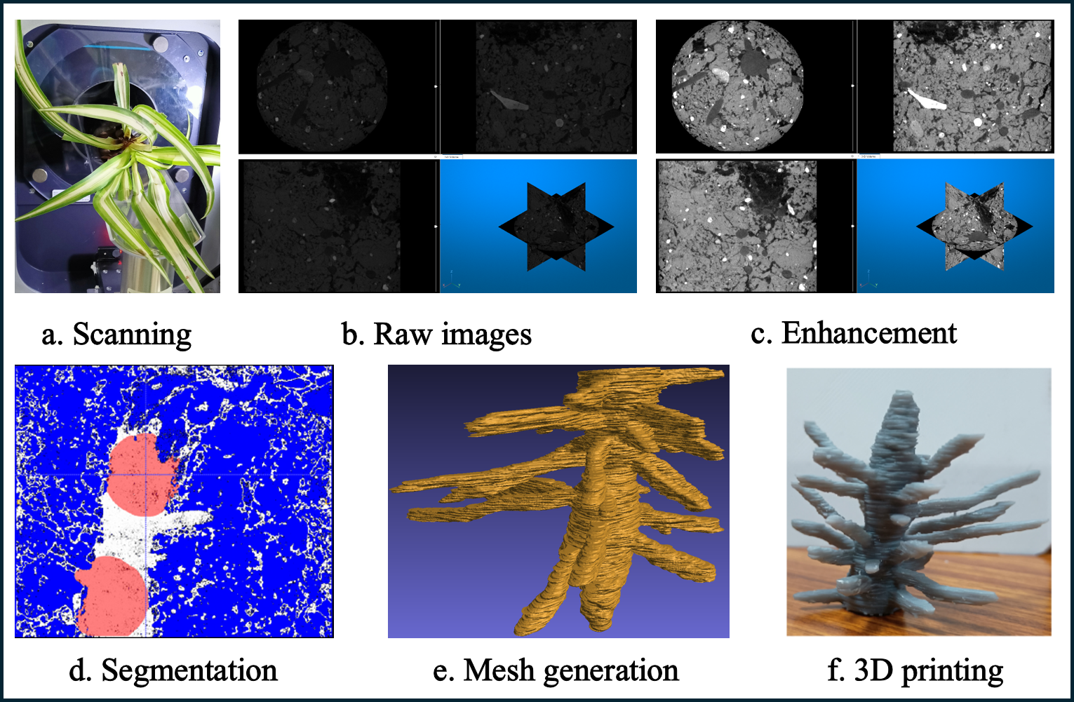
The figure here illustrates the reconstruction pipeline applied to the roots of a chlorophyta plant, that was scanned using a micro-CT scanner and recontructed using region growing segmentation algorithm and printed using Form 3+ SLA 3D printer
3D Model from Light-Based Scanners
Light-based scanning allows non-invasive 3D capture of surface features for prosthetic design, digital archiving, and reverse engineering. CAD models from structured light scans offer significant insight in personalised product development and AI-based analysis.
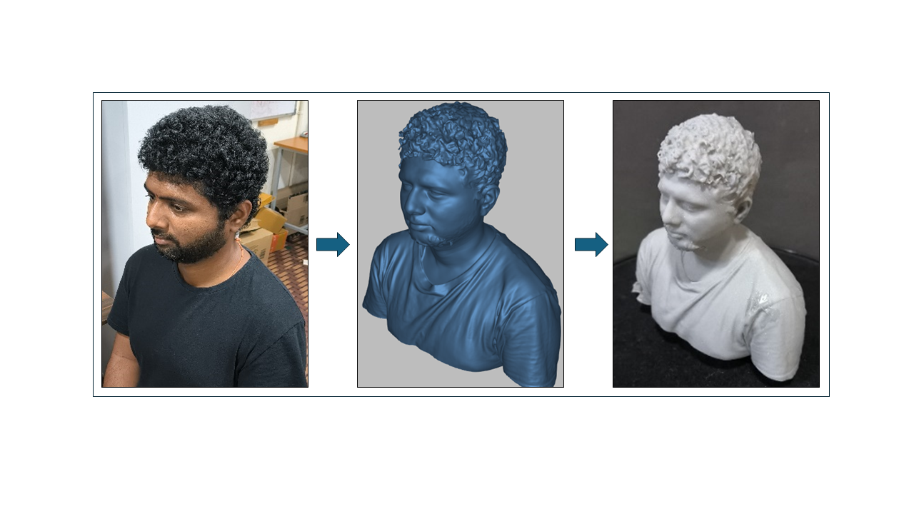
The image above shows the stages of structured light pattern-based reconstruction applied to a human face model. The video demonstrates the use of the Artec Eva scanner, hosted at BioM3D lab, in scanning and creating a geometric model of a human bust.
Mesh Smoothing
Mesh smoothing is a critical post-processing step applied to all tessellations. It removes staircase artefacts and jaggedness introduced by imaging and hardware limitations, producing smooth continuous geometries.
The trade-off: smoothing can inadvertently erode intricate features. A spectral stopping criterion using the Laplace-Beltrami eigenvalue spectrum was developed to address this. The video demonstrates the effect of Laplacian smoothing with a sufficient scaling factor on the geometry of a coronary artery.
Morphological Extraction
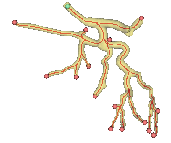
Post-thresholding morphological filters (erosion, dilation, etc.) refine binary masks to extract features like branching, length, and continuity from complex biological shapes such as plant roots and blood vessels. A desktop PyQt-based application was built for batch morphological analysis of vascular networks.
3D Printing
Using STL models derived from imaging, prototypes are printed via SLA/FDM processes. 3D prints enable physical validation, educational demonstrations, and pre-surgical rehearsals.
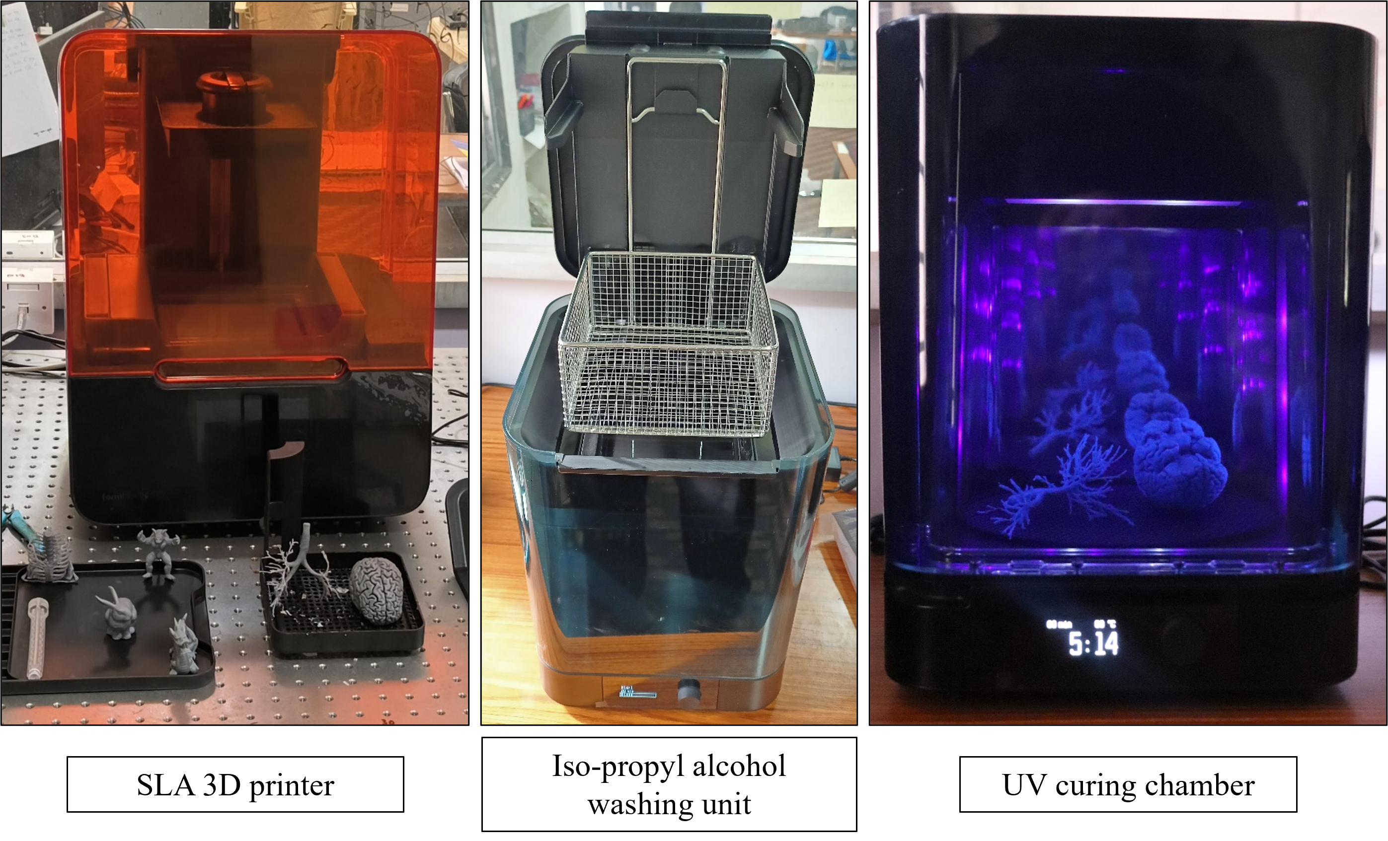
The image shows the Formlabs Form 3+ SLA 3D printing facility, along with washing and curing units at BioM3D lab. The video shows printing of a flexible tube using a material with Shore hardness of 80A.